Scoliosis X-Ray: When You Need One and What Happens Next

If you have noticed a shoulder sitting higher, a rib area that sticks out more when you bend forward, or a teenager who suddenly looks tilted in photos, the uncertainty can escalate quickly. You start wondering if it is posture, growth, a heavy school bag, or something structural. Then you hit the next step question: Do we need a scoliosis X-ray, or is that overkill?
This guide is here to make that decision calmer. You will learn when imaging is actually useful, what it can answer, how results are measured, and what a sensible next step looks like for teens, adults, and parents in Hong Kong who want clarity, not drama.
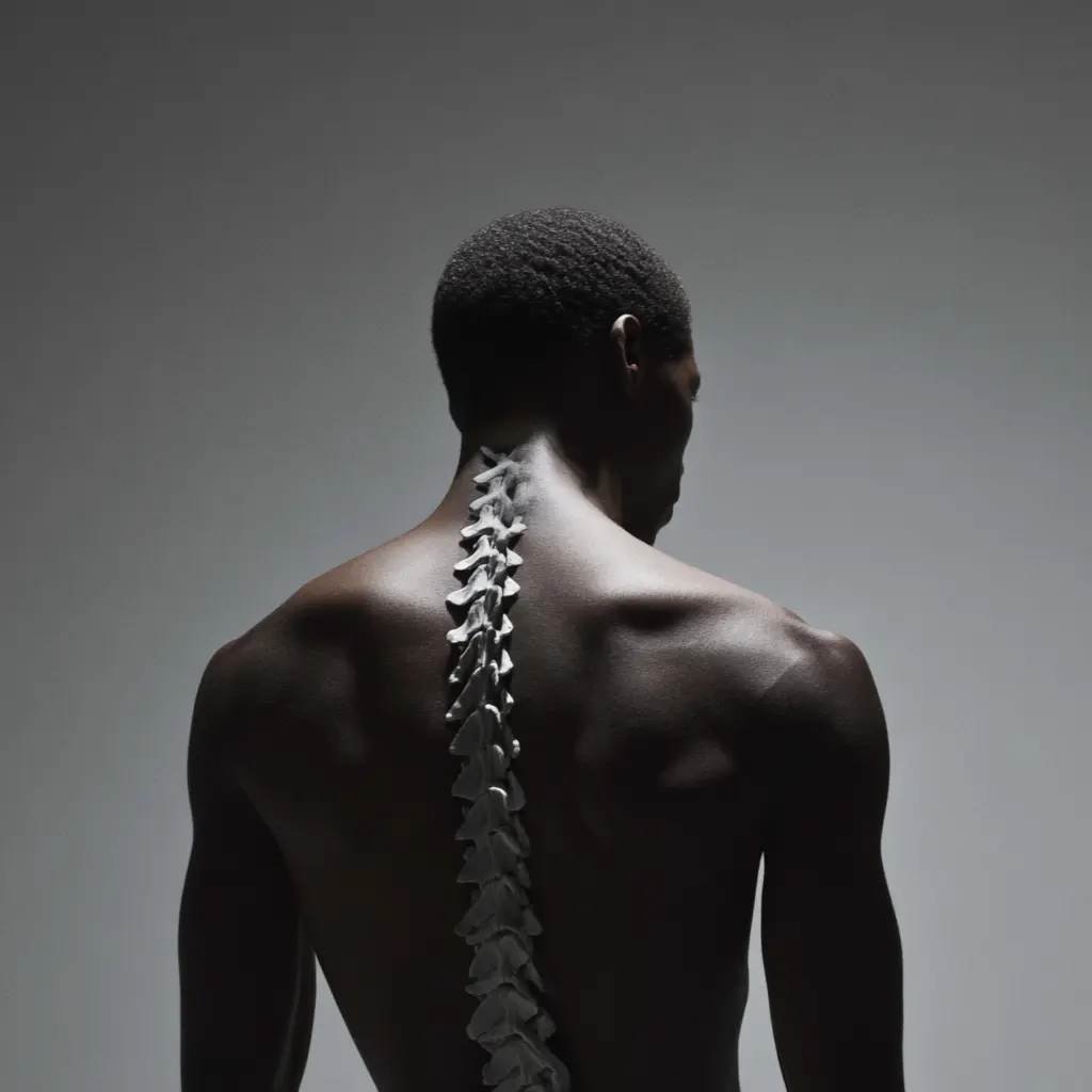
One useful anchor before we start: Scoliosis is typically defined as a sideways curve of 10 degrees or more on X-ray, often with a rotational component, and it is common enough that you are not the only family navigating it. Adolescent idiopathic scoliosis is commonly reported in about 1% to 3% of adolescents.
When people hear rotation, it simply means the vertebrae can twist slightly, which can make one side of the ribs look more prominent when bending forward.
When a Scoliosis X-Ray Is Actually Recommended
The most frustrating part is that scoliosis rarely announces itself with a clear alarm. It shows up as small visual changes, vague discomfort, or a parent noticing a change before the teen feels anything at all. That is why a good process matters.
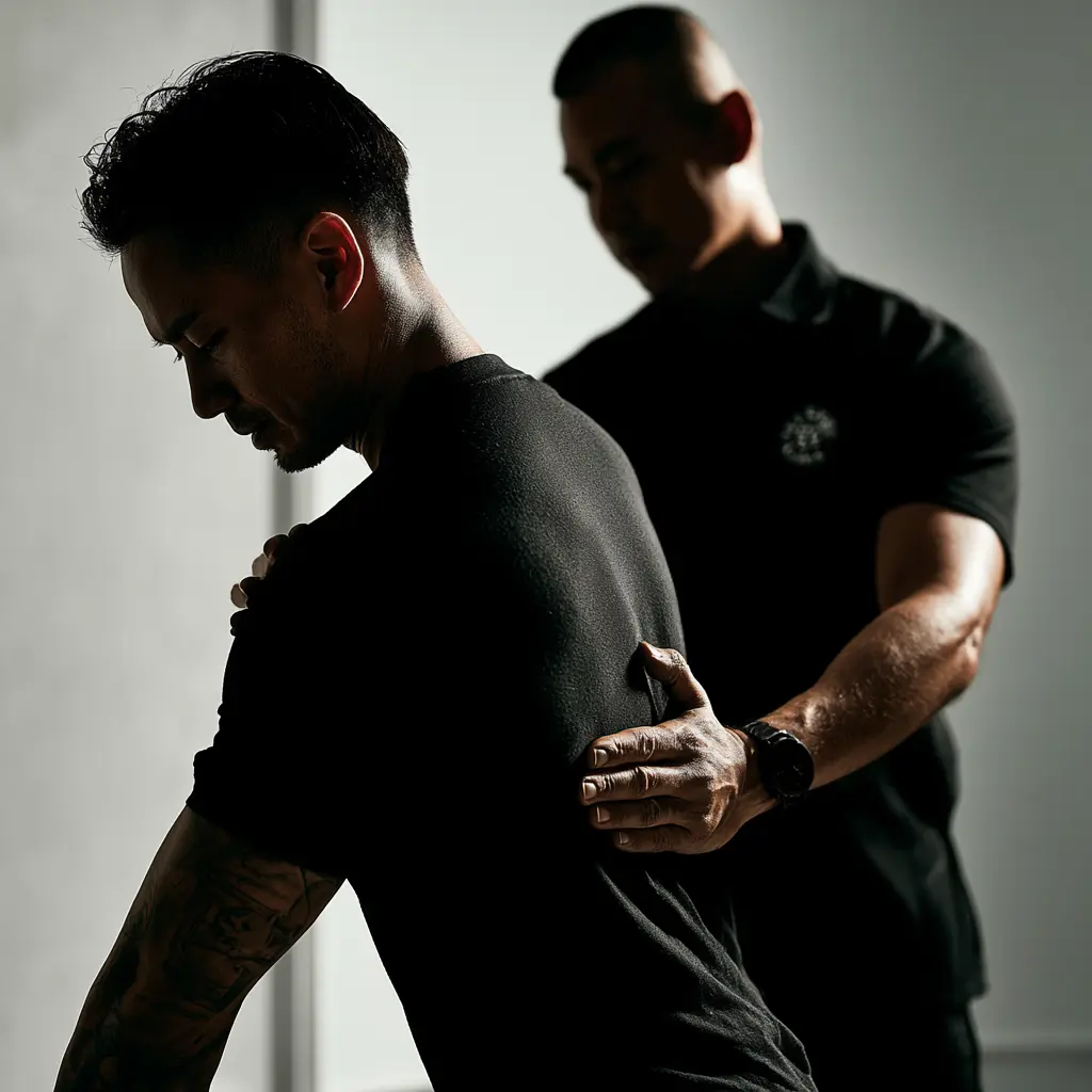
Signs and Symptoms That Prompt an X-Ray Referral
A scoliosis X-ray is commonly considered when a physical exam suggests a structural curve, especially if you are seeing consistent asymmetry, such as:
- Uneven shoulders or shoulder blades
- A visible waistline shift or one hip appearing higher
- Rib prominence when bending forward
- Clothing that suddenly hangs unevenly
- Back discomfort that keeps returning, especially with growth or training load
An exam can raise suspicion, but a standing spinal X-ray is what confirms the curve and allows it to be measured accurately.
Why Imaging Is Not Always the First Step
Here is the part people rarely hear: you do not need an X-ray every time someone looks slightly uneven.
Some asymmetry is normal. Some is driven by position, muscle tone, or habit. Especially in teens, a structured physical exam can clarify whether the spine looks like it is adapting temporarily or whether it behaves like a fixed structural curve.
Age, Growth, and History Factors That Influence Imaging Decisions
This is where decision-making matters most.
In teens, growth is the main wildcard. The same curve behaves differently depending on how much growth is left. In adults, imaging is more often guided by symptoms, function, and whether the curve appears stable over time.
Two helpful questions that guide the decision:
Is this likely a structural curve, or does it behave more like posture and positioning?
If it is structural, is this a situation to measure and monitor, or is the pattern more likely to change during growth or with time?
The goal is not to chase numbers. The goal is to decide what the numbers mean for real life: sleep, energy, comfort, training, school days, and confidence.
What Imaging Shows (And What It Doesn't)
A lot of people expect an X-ray to deliver a neat answer. Mild. Moderate. Severe. Done. In reality, an X-ray is more like a map. It shows structure clearly, but you still need to interpret what the map means for the person standing in front of you.
A practical detail many people want to know: scoliosis imaging is usually done as standing full-spine views, so the curve is measured under normal load. This matters when you are comparing images over time.
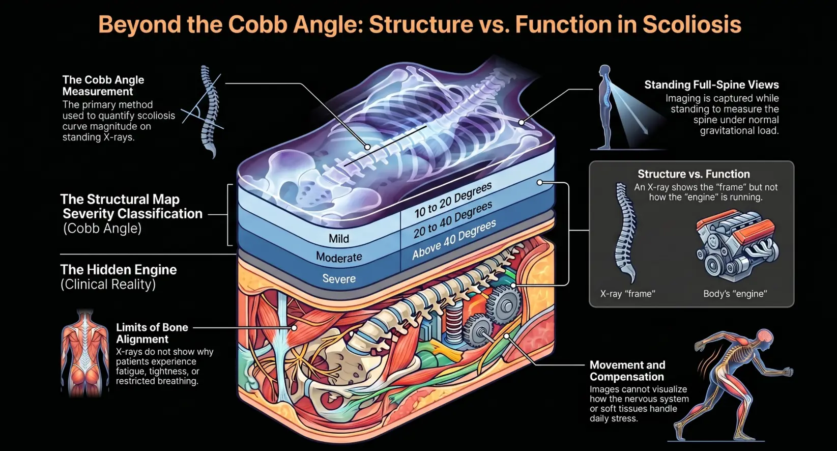
How X-Ray Images Are Used to Measure Curve Severity
The key measurement is the Cobb angle. It is the most widely used way to quantify curve magnitude on plain radiographs, and Radiopaedia's explainer on the Cobb angle is a helpful reference.
A simple framework many reports use is:
- Mild: roughly 10 to 20 degrees
- Moderate: roughly 20 to 40 degrees
- Severe: above 40 degrees
This framework helps guide monitoring and referral decisions, but it does not replace clinical judgment. In plain terms, mild often means the immediate focus is observation and function first, especially if day-to-day symptoms are manageable.
Limits of an X-Ray Without Clinical Assessment
An X-ray shows bone alignment. It does not tell you why your upper back feels tight at the end of the day, why sitting triggers fatigue, or why one side of the ribs feels restricted when breathing.
You can have a measurable curve and feel fine. You can also have a smaller curve and feel very limited. That is why the useful question is not what number is it, but what does this mean for movement, load tolerance, and daily life.
What the Image Cannot Show in Soft Tissue and Function
X-rays do not show soft tissue health or how your nervous system is responding. They cannot show how you move, how you compensate, or how your body handles stress.
Plain language version: an X-ray can show the shape of the frame. It cannot show how the engine is running.
That is where a posture and structural assessment adds what the image alone cannot provide.
How Results Fit Into a Full Assessment Process
A lot of clinics treat imaging like the finish line. At ATLAS Chiropractor in Hong Kong it is one data point inside a larger system.
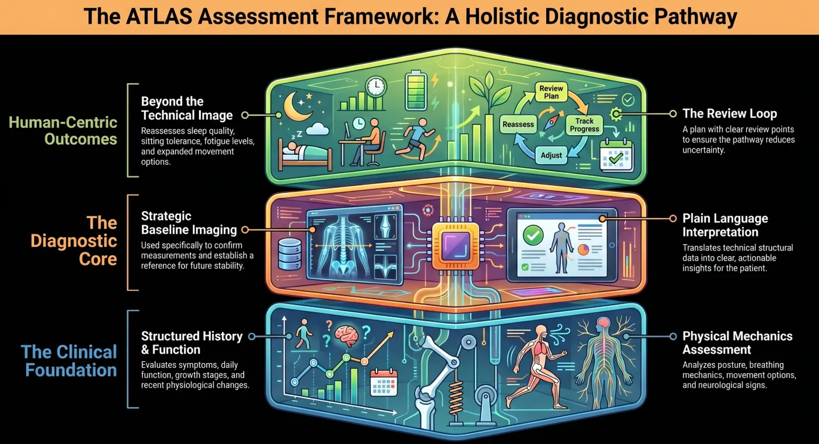
End-to-End Sequence: Assessment, Imaging, Interpretation, Plan, Review
A clear sequence reduces uncertainty.
A sensible pathway looks like this:
- Structured history covering symptoms, daily function, growth stage, and recent changes
- Physical assessment of posture, movement options, breathing mechanics, and neurological signs — part of our broader paediatric chiropractic assessment process for growing teens
- Imaging only when indicated to confirm and measure the curve
- Interpretation in plain language
- A plan with clear review points
This is the practical meaning of we assess, we don't guess.
Using Results to Establish a Clear Baseline
When imaging is indicated, its biggest value is often the baseline. A baseline makes future decisions clearer.
It helps answer whether the curve is stable, whether growth may influence change, and whether symptoms match the structural picture.
What Follow-Up Reassesses Besides the Image
Follow-up is not only about the curve.
It also looks at whether sleep improves, sitting tolerance increases, training feels steadier, fatigue changes, and movement options expand. This is how assessment stays human instead of purely technical.
After Imaging: Understanding Your Next Steps
This is the moment most people get stuck. They have the report. They have a number. Now what?
A helpful way to think about it is that teens and adults often need different next steps even with the same Cobb angle. Teens are managed with growth in mind. Adults are managed with function and stability in mind.
What Happens if the Report Shows a Mild Curve
For many teens and adults, mild curves are managed with observation and structured guidance.
This often includes a monitoring plan, targeted exercise, realistic posture education, and support for daily activities.
A key reassurance: many people do not progress into severe categories.
What Changes When the Curve Is More Likely to Change Over Time
Progression risk simply means that follow-up needs to be more deliberate.
In growing adolescents, monitoring intervals are often closer during faster growth phases. Clinical guidance commonly references re-checks during growth to track change, including the approach described in Mayo Clinic's scoliosis guidance.
What Monitoring Typically Involves Over Time, and When Imaging Is Repeated
Monitoring is not just come back someday.
It means agreed review points where function is reassessed, and imaging is repeated only if there is a clear reason to do so. Those reviews typically look at visible posture markers, symptom patterns, daily function, growth stage, and tolerance for school, sport, training, or desk work.
For many growing teens, repeat X-rays are commonly discussed in practical intervals, such as every 6 months when monitoring is clinically indicated.
The aim is to always know what you are watching and what would trigger the next step.
Is an X-Ray Safe for Teenagers?
A scoliosis X-ray uses a low dose of radiation, and for most teenagers, the risk from a single scan is considered very small. The bigger concern is repeat imaging during growth, because monitoring can involve follow-up scans over months or years. That is why clinicians only order X-rays when the result will change the plan, and why most imaging is done using modern low-dose techniques and careful positioning.
If you are concerned, ask the imaging centre whether they use low-dose protocols for scoliosis and how often repeat images would be recommended based on growth stage and the findings. The goal is simple: get the information you need, without scanning more than necessary.
How Often Should an X-Ray Be Repeated?
There is no single schedule, because it depends on growth stage, curve size, and whether a change is suspected. For teens who are still growing, follow-up X-rays are often spaced out over months rather than weeks, and may be repeated periodically during faster growth phases to check whether the curve is changing.
For adults, imaging is usually less frequent and is typically repeated only if symptoms, function, or clinical findings change. The safety goal is to get the information needed to guide decisions while avoiding unnecessary repeat scans, so imaging should be planned around clear review points rather than done routinely just in case.
Can Scoliosis Be Assessed Without an X-Ray?
Yes, to a point. A structured history and physical exam can screen for scoliosis, identify red flags, and help decide whether imaging is likely to change the plan. However, confirming scoliosis and accurately measuring the curve usually requires an X-ray because that is how the Cobb angle is measured.
If safety is your concern, the key is that X-rays are typically used when the result will guide monitoring or care decisions, not as a routine scan for every mild asymmetry. The goal is to get a clear baseline when needed, then limit repeat imaging to planned review points.
Sources
Mayo Clinic — Scoliosis: Diagnosis and treatment. Retrieved from https://www.mayoclinic.org/diseases-conditions/scoliosis/diagnosis-treatment/drc-20350721
NCBI Bookshelf / StatPearls — Adolescent Idiopathic Scoliosis. Retrieved from https://www.ncbi.nlm.nih.gov/books/NBK499908/
NCBI Bookshelf / StatPearls — Scoliosis. Retrieved from https://www.ncbi.nlm.nih.gov/books/NBK493367/
Final Thoughts
If you are searching for scoliosis xray, you are usually trying to decide what to do next without overreacting.
The grounded approach is this: assessment first, imaging when it adds clarity, and follow-up that is planned rather than reactive. At ATLAS, that is exactly what we mean by we assess, we don't guess.
If you are in Hong Kong, the practical path is usually to start with a structured clinical assessment and then arrange imaging when it is likely to change the decision, rather than scanning first and hoping the report explains everything.
ATLAS guides clients through a structured assessment process, explains findings in everyday language, and maps out follow-up so you know exactly what you are watching and why. If you are ready for a calm, precise next step, book an evaluation with ATLAS.